Musculoskeletal X–rays for Medical Students
Samenvatting
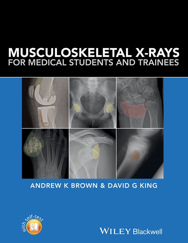
Musculoskeletal X–rays for Medical Students provides the key principles and skills needed for the assessment of normal and abnormal musculoskeletal radiographs. With a focus on concise information and clear visual presentation, it uses a unique colour overlay system to clearly present abnormalities.
Musculoskeletal X–rays for Medical Students:
Presents each radiograph twice, side by side once as would be seen in a clinical setting and again with clearly highlighted anatomy or pathology
Focuses on radiographic appearances and abnormalities seen in common clinical presentations, highlighting key learning points relevant to each condition
Covers introductory principles, normal anatomy and common pathologies, in addition to disease–specific sections covering adult and paediatric practice
Includes self–assessment to test knowledge and presentation techniques
Musculoskeletal X–rays for Medical Students is designed for medical students, junior doctors, nurses and radiographers, and is ideal for both study and clinical reference.
Specificaties
Inhoudsopgave
<p>Acknowledgements, viii</p>
<p>Part 1: Introduction, 1</p>
<p>1 Musculoskeletal X –rays, 3</p>
<p>Introduction, 3</p>
<p>Basic principles of requesting plain radiographs of bones and joints, 4</p>
<p>Basic principles of examining and reporting plain radiographs of bones and joints, 6</p>
<p>Normal anatomy on musculoskeletal X–rays, 8</p>
<p>Part 2: Pathology, 21</p>
<p>2 Trauma, 23</p>
<p>Bone and joint injuries, 23</p>
<p>Specific injuries, 41</p>
<p>Spine, 58</p>
<p>Paediatric fractures, 67</p>
<p>Fractures in child abuse, 73</p>
<p>Further reading, 76</p>
<p>3 Arthritis, 77</p>
<p>Osteoarthritis, 77</p>
<p>Rheumatoid arthritis, 80</p>
<p>Crystal arthropathy, 83</p>
<p>Gout, 83</p>
<p>Calcium pyrophosphate disease, 86</p>
<p>Psoriatic arthritis, 91</p>
<p>Axial spondyloarthritis (ankylosing spondylitis), 95</p>
<p>4 Tumours and tumour –like lesions, 98</p>
<p>Radiological evaluation of the patient, 98</p>
<p>X–rays general principles, 101</p>
<p>Malignant tumours, 105</p>
<p>Bone metastases, 105</p>
<p>Multiple myeloma, 107</p>
<p>Plasmacytoma, 108</p>
<p>Osteosarcoma, 110</p>
<p>Chondrosarcoma, 111</p>
<p>Ewing s sarcoma, 113</p>
<p>Benign tumours, 114</p>
<p>Exostosis (Osteochondroma), 115</p>
<p>Osteoid osteoma, 116</p>
<p>Tumour –like lesions, 117</p>
<p>Simple bone cyst, 118</p>
<p>Infection, 119</p>
<p>5 Metabolic bone disease, 120</p>
<p>Osteoporosis, 120</p>
<p>Osteomalacia, 121</p>
<p>Hyperparathyroidism, 122</p>
<p>Chronic kidney disease metabolic bone disorder, 124</p>
<p>Haemochromatosis, 126</p>
<p>6 Infection, 128</p>
<p>Routes of spread, 128</p>
<p>Causative organisms, 128</p>
<p>Osteomyelitis, 129</p>
<p>Septic arthritis, 134</p>
<p>Infective discitis, 136</p>
<p>7 Non –traumatic paediatric conditions, 138</p>
<p>Developmental dysplasia of the hip, 138</p>
<p>Perthes disease, 140</p>
<p>Tarsal coalition, 141</p>
<p>Osteochondritis dissecans, 143</p>
<p>8 Other bone pathology, 144</p>
<p>Paget s disease of bone, 144</p>
<p>Hypertrophic Osteoarthropathy (HOA), 147</p>
<p>Avascular necrosis, 147</p>
<p>9 Joint replacement, 149</p>
<p>Hardware failure and aseptic loosening, 149</p>
<p>Infection, 154</p>
<p>Malalignment and instability, 155</p>
<p>Periprosthetic fracture, 156</p>
<p>Part 3, 157</p>
<p>Self–assessment questions, 159</p>
<p>Self–assessment answers, 172</p>
<p>Index, 185</p>