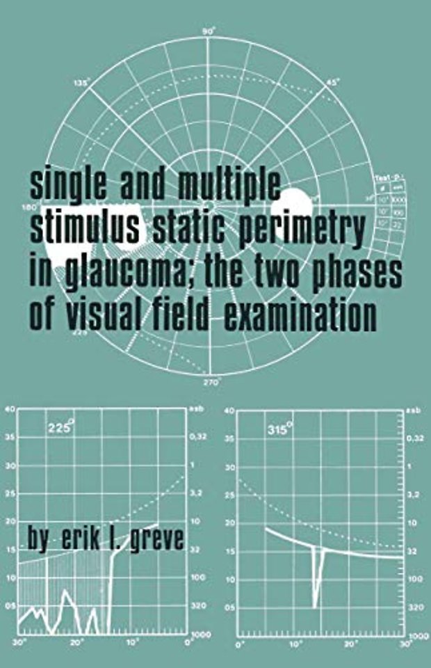
Single and Multiple Stimulus Static Perimetry in Glaucoma; The Two Phases of Perimetry
The Two Phases of Perimetry
Samenvatting
does not vary more than 1 % over the whole surface. Stimulus: The luminance of the stimuli can be regulated by means of neutral density filters. These filters are neither entirely neutral nor completely uniform, but these variations are not significant for perimetry. The maximum luminance of the stimuli is standardized at 1000 asb (316 2 cd.m-). The standard GP for kinetic perimetry has 3 neutral density filters which reduce the luminance by 0.5, 1.0 and 1.5 log units respectively. The modification of the GP for static perimetry is supplied with an additional series of four neutral density filters, which allow luminance steps of 0.1, 0.2, 0.3 and 0.4 log units. The resulting luminance values, expressed in apostilb, are given in Table I. TABLE I Luminance values of the Goldmann perimeter 4 3 2 steps lux 1430 450 143 45 3,1 asb 1000 315 100 31,5 3,1 2 cd/m 315 100 31,5 10 3,1 ßL ±30 10 3 3,1 L log L asb 3 2,5 2 2 1,5 0,5 2 Background: 31,5 asb. = 10 cd/m • Coefficient of reflection 0,7. 1 2 (1 asb = -cd/m).